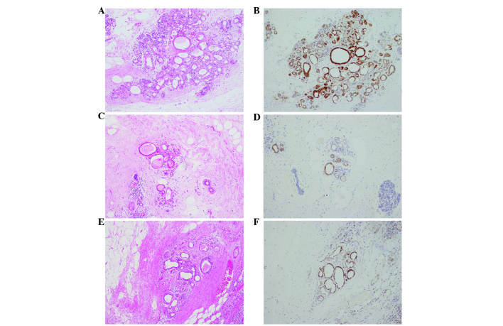
Figure 6.
Immunohistochemical staining of the surgical specimens counterstained with hematoxylin and eosin (HE). Oncocytic cells were observed in the peritumoral lesion, which also showed reactivity for anti-mitochondrial antibody. (A) Right upper outer quadrant. HE staining. (B) Right upper outer quadrant. Mitochondrial staining. (C) Right outer quadrant. HE staining. (D) Right outer quadrant. Mitochondrial staining. (E) Left upper inner quadrant. HE staining. (F) Left upper inner quadrant. Mitochondrial staining. Magnification, ×100.

