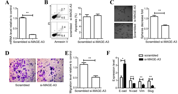
Figure 3.
Effect of MAGE-A3 knockdown on A549 cells. (A) Evaluation of efficiency of si-mediated MAGE-A3 knockdown via qRT-PCR analysis. (B) Effect of MAGE-A3 knockdown on cell apoptosis was detected through flow cytometry and quantified. (C) Depletion of MAGE-A3 inhibited cell colony formation. (D) Representative distinction of migration in A549 cells treated by scrambled and specific MAGE-A3 siRNA. (E) Downregulation of MAGE-A3 in A549 cells resulted in reduced cell migration. (F) Effect of MAGE-A3 knockdown on the expression of epithelial-mesenchymal transition markers was detected by qRT-PCR. All experiments were repeated ≥3 times. Data were compared using paired two-tailed t-tests. *P<0.05, **P<0.01, ***P<0.001 vs. the scrambled control group. MAGE, melanoma-associated antigen; si, small interfering RNA; EMT, epithelial-mesenchymal-transition marker; E-cad, epithelial-cadherin; N-cad, neural-cadherin; SLUG, snail family transcriptional repressor 2; Vim, vimentin; bp, base pairs; qPCR, quantitative polymerase chain reaction.

