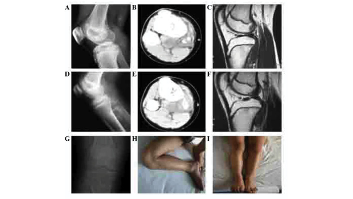
Figure 3.
Case 2: Imaging examinations, including (A, D and G) X-ray, (B and E) computed tomography and (C and E) magnetic resonance imaging (MRI). (A-C) A space-occupying lesion in the proximal region of the right tibia, with no intramedullary involvement. (D-F) Radiological changes owing to pre-operative chemotherapy; the mass was well-defined and calcification was detected within it. MRI indicated a smaller volume of tumor. (G) X-ray at the end of 108 months of follow-up showing no local recurrence. (H and I) Normal limb function, as demonstrated by as demonstrated by normal (H) flexion and (I) extension, at the end of the follow-up period.

