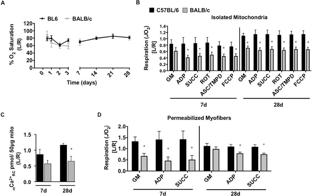
Figure 3. Limb muscle mitochondrial response to sub-acute hindlimb ischemia.
Sub-acute femoral artery occlusion was performed on C57BL/6 and BALB/c mice by placement of a single AC (1AC) on the proximal portion of the femoral artery, immediately proximal to the epigastric arterial branch. Paw tissue oxygen saturation (%SO2), measured via white light reflectance spectroscopy, was analyzed to determine distal limb O2 (A). B. Mitochondrial respiration (oxygen consumption rate-JO2, as a measure of electron transport system flux) was measured in isolated mitochondria using high-resolution respirometry. The respiratory states assessed were: 1.) glutamate/malate supported state 2 respiration (GM) 2.) glutamate/malate supported state 3 (ADP) 3.) GM and succinate supported state 3 (SUCC) 4.) succinate supported state 3 (ROT) 5.) ascorbate/TMPD supported state 3 (ASC/TMPD) and 6.) FCCP supported state 3 uncoupled respiration (FCCP). C. Ca2+ reuptake was measured in ischemic (L) and control (R) mitochondria isolated from the limb plantar flexors (gastrocnemius, plantaris, and soleus) as a function of mitochondrial integrity and resistance to permeability transition. D. Oxygen consumption was also determined in saponin permeabilized myofiber bundles isolated from the gastrocnemius muscle. Data are representative of the ratio of the ischemic limb (L) to the untreated control limb (R). * P<0.05 vs. C57BL/6. All data are means ± SEM and representative of N≥6/strain/time point.

