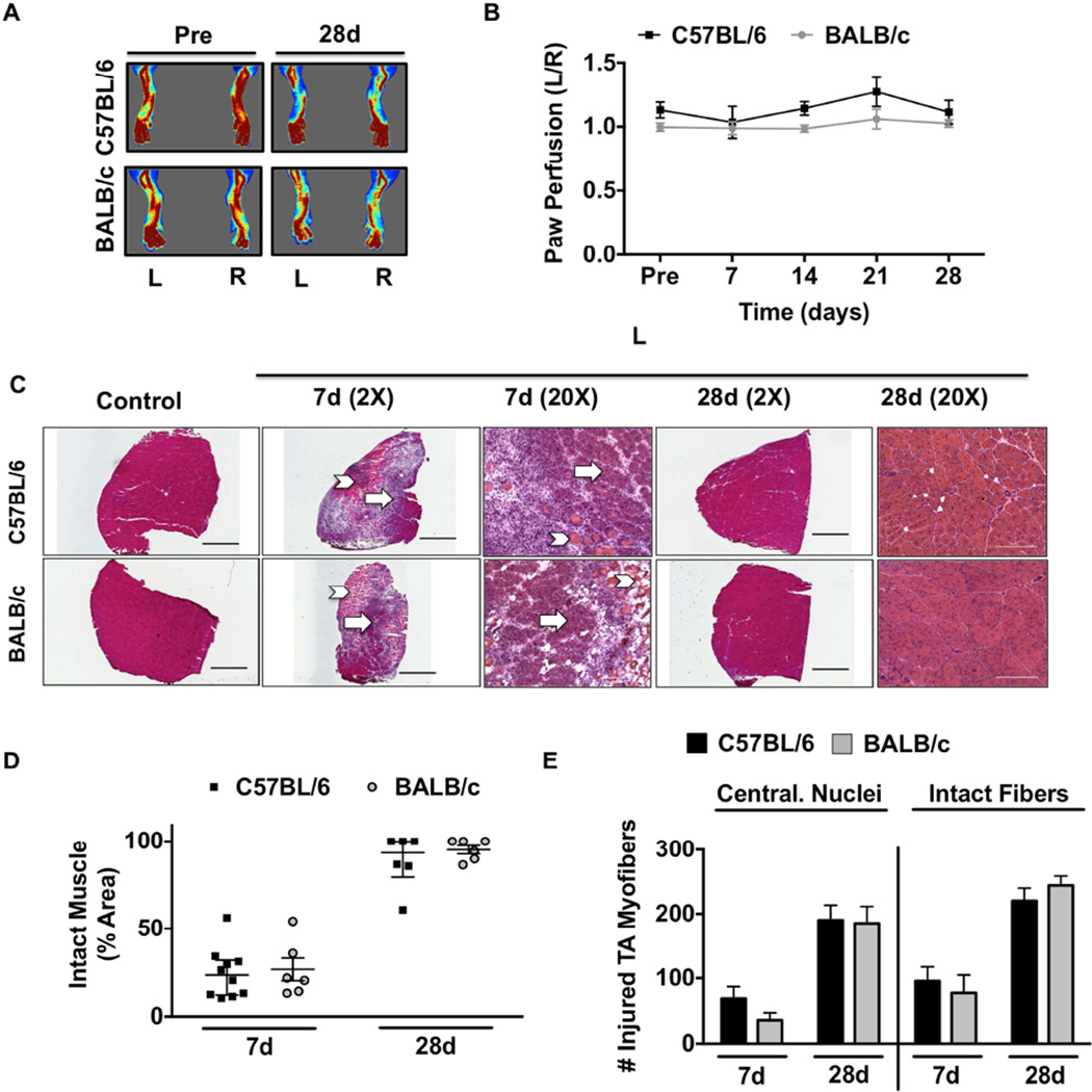
Figure 4. Strain independent myopathy with non-ischemic myotoxin injury.
Cardiotoxin (CTX) was injected intramuscularly into the TA and the lateral/medial heads of the gastrocnemius muscle. A sham injection (Ctrl) of equal volume sterile saline was injected in the same muscles in the contralateral control limb (R). A. Representative laser Doppler perfusion images (LDPI) of the paw before and during recovery from cardiotoxin injection. B. Quantification of blood flow in the hind paw, presented as the ratio of the CTX injected (L) limb to the sham injected control (R) limb. C. Representative 2X and 20X images of hematoxylin and eosin (H&E) stained sections of tibialis anterior (TA) muscles 7 and 28 days after CTX (L) injury. Arrows indicate intact muscle; Chevrons indicate regions of anucleate necrotic fibers. Scale bar inlays are 1000µm in length(2X), and 200µm in length(20X). D. Muscle regeneration was quantified from 2X H&E images by measuring the total regions of interest (ROI; area) containing necrotic/anuclueate myofibers, which is represented by median and interquartile range. E. Total myofibers with centralized nuclei (Central. Nuclei) and total myofiber number (Intact Fibers) were determined in representative 10X images. All data are means ± SEM and representative of N≥6/strain/time point.

