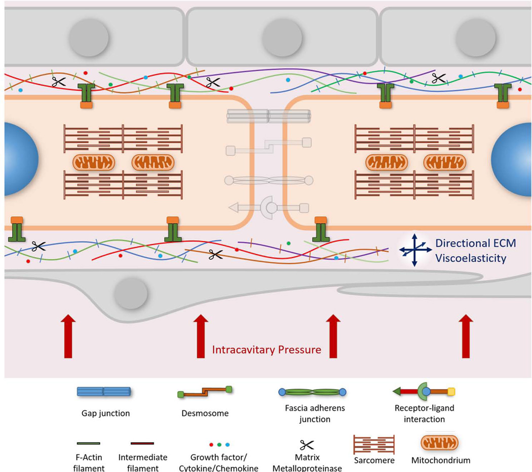
Figure 3. Cell-ECM Interactions in the Myocardium.
Cardiac cells are surrounded by the extracellular matrix (ECM). It is the anchoring source that provides structural as well as molecular support to cardiac cells. The ECM is composed of a wide variety of micro-and macromolecules including fibrous proteins, proteoglycans and incorporates growth factors, cytokines and chemokines. As such, the ECM provides important biochemical and biophysical signaling cues to cardiac myocytes. The balance between synthesis and degradation of the ECM is maintained by matrix metalloproteinases. Cardiac myocytes connect to the ECM at costamere complexes. Costameres are mechanotransducing complexes that mainly link Z-discs laterally to the ECM and consist of integrin and dystrophin-glycoprotein complexes. Integrins play a critical role in forming focal adhesions in vitro that couple actin filaments through linker proteins to the ECM. At costameres the incoming mechanical load regulates biochemical signaling pathways. Cytoskeletal organization and cell geometry are significantly influenced by how the ECM is presented to the cell. As a result dysregulation of the ECM may lead to cytoskeletal disarray and aberrant cell shape. The viscoelastic properties of the ECM also influence the contractile properties of the myocardium. In addition, the intracavitary pressure of the native heart is transmitted through the ECM and acts as a major determinant of the biomechanical forces exerted on myocardial cells. Aspects of cell-cell interactions have been deemphasized to reduce visual clutter.

