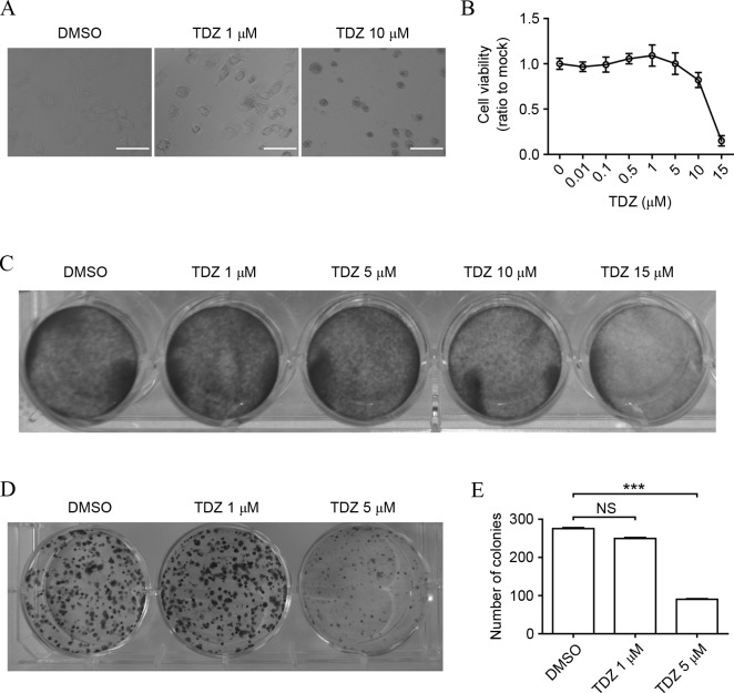
Figure 1.
TDZ exhibits cytotoxicity in lung CSCs. (A) Morphology of A549 sphere cells altered following treatment with 1 or 10 µM TDZ for 48 h. Scale bar, 200 µm. (B) Cell viability of A549 sphere cells decreased in a dose-dependent manner following TDZ treatment (0.01, 0.1, 0.5, 1, 5, 10 or 15 µM) for 48 h. Cell viability was determined by MTT assay (C) TDZ attenuated A549 sphere cell viability. A549 sphere cells were treated with TDZ (1, 5, 10 or 15 µM) for 48 h and then subjected to crystal violet staining. (D) TDZ-treated A549 sphere cells formed fewer colonies, as shown by crystal violet staining. (E) Statistical analysis of colony formation assay. The y-axis indicated the total number of colonies in 1 well. All experiments were repeated three times with DMSO as a control. All data shown are expressed as the mean ± standard deviation (n=3). ***P<0.001. TDZ, thioridazine; DMSO, dimethyl sulfoxide; NS, no significance.

