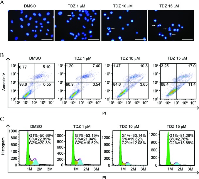
Figure 2.
TDZ induced apoptosis and cell cycle arrest in A549 sphere cells. (A) Increased nucleic fragmentation (arrow) was observed in A549 sphere cells following 2-day treatment with TDZ (1, 10 or 15 µM), as detected by Hoechst staining. Scale bar, 200 µm. (B) Apoptosis was detected in TDZ-treated (1, 10 or 15 µM) A549 sphere cells by Annexin V/PI double staining with fluorescence-activated cell sorting analysis. (C) G1-phase cell proportion increased with concentration of TDZ treatment (1, 10 or 15 µM) on A549 sphere cells. The areas of green, yellow and blue equaled the percentage of cells in G1, S and G2 phase, respectively. All experiments were repeated three times with DMSO as a control. TDZ, thioridazine; PI, propidium iodide; DMSO, dimethyl sulfoxide.

