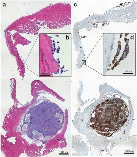
Fig. 1.

Tissue sections of the abdominal wall from a reference animal treated with unlabeled MX35 visualizing a macroscopic tumor (bottom of a and c) and microscopic tumors (b and d). a and b are stained with H&E while c and d show the dense distribution (in brown) of the MX35 antigen on tumor cells using IHC
