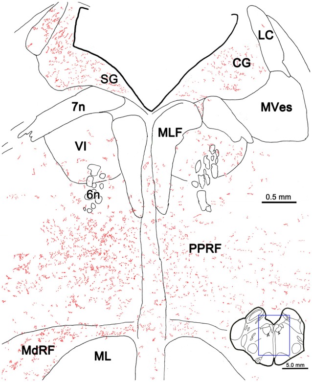
Figure 5.

PhaL labeled reticuloreticular axons at the level of the abducens nucleus. The pattern of reticuloreticular axons (lines) and terminals (stipple) is illustrated for the region shown by a box in the inset. The injection site is illustrated in Figure 4. Note the ipsilateral predominance of the terminal labeling in the PRF and MdRF.
