. 2017 Apr 21;26(2):104–112. doi: 10.5607/en.2017.26.2.104

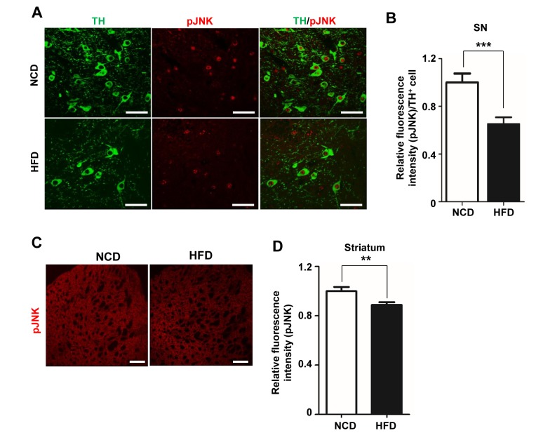
Fig. 3. The decrease in TH is associated with a reduction in phosphorylated JNK levels. (A) Brain slices (25 µm thick) were double-immunofluorescence stained by incubating overnight with anti-TH and rabbit anti-pJNK antibodies (1:500). TH (green) and pJNK (red) fluorescence in the SN were visualized by confocal microscopy. (B) Relative intensity of pJNK fluorescence per TH-positive cell in the SN was measured using ImageJ software (n=10 slides per condition, n=6/group). (C) pJNK immunoreactivity was detected in the striatum. (D) The intensity of pJNK fluorescence in the striatum was measured and graphed. Scale bars: 50 µm. *p<0.05, **p<0.01.

Fig. 3