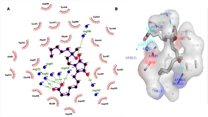
FIGURE 5.

(A) 2D, and 3D (B) representation of docked structure of COI1 generated by Ligplot and PyMol tool; The 2D representation of 3D structure depicted H-bond interaction of ZINC27640214 with COI1 amino acid residues, ARG85, ARG121, ARG349, ARG410, and ARG500 (green line) whereas the amino acid residues such as ARG85, ALA86, MET88, PHE89, LEU91, ARG349, TYR387, ARG410, VAL412, ALA444, TYR446, LEU471, GLU498, ARG500, and TRP523 were interacted through hydrophobic bonding (in red color).
