Figure 3.
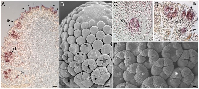
Morphology of Anacyclus capitulum and florets, and tissue-specific expression of AcCYC2 genes during capitulum development. (A) Section of a Anacyclus capitulum hybridized with a digoxigenin-labeled probe complementary to AcCYC2d. Youngest flower meristems (fm) can be seen at the top, older flowers to the left and right. AcCYC2d transcripts accumulate in young flower meristems and in meristems that are initiating corolla lobe primordia (*). In older flowers mRNA is detectable in developing stamens (st) and ovules (ov). No signal is detectable in developing corolla lobes (lb). (B) Scanning electron microscopy (SEM) image of a capitulum of an age comparable to that in (A). (C) Close up of an ovule displaying AcCYC2d expression. (D) Young disc floret showing AcCYC2a mRNA signal in stamens and ovule but not in corolla lobes. (E) SEM image of disc flowers comparable to those in (D).

