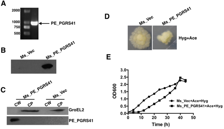
Figure 1. The effect of PE_PGRS41 on the growth of M. smegmatis.
(A) PCR amplification of PE_PGRS41 encoding sequence from M. tuberculosis genome approximately 1086 bp. (B) M. tuberculosis PE_PGRS41 was expressed in M. smegmatis and detected using Western blotting. Cell lysates of Ms_Vec and Ms_PE_PGRS41 were subjected to Western blot to determine the expression of His-tagged PE_PGRS41 protein in M. smegmatis by anti-His antibody. (C) Cell fractionation experiments were performed to determine the sub-cellular localization of PE_PGRS41, GroEL2 protein serves as a cytoplasm marker of M. smegmatis, CW represents cell wall; CP represents cytoplasm. (D) The morphology of Ms_Vec and Ms_PE_PGRS41 were detected after induction by acetamide in the presence of hygromycin. (E) Ms_Vec and Ms_PE_PGRS41 were grown in Middlebrook 7H9 medium supplemented with 0.05% Tween 80, 1% acetamide and 0.2% glycerinum, with hygromycin (100 μg/ml). The OD600 was determined at an interval of 4 h.

