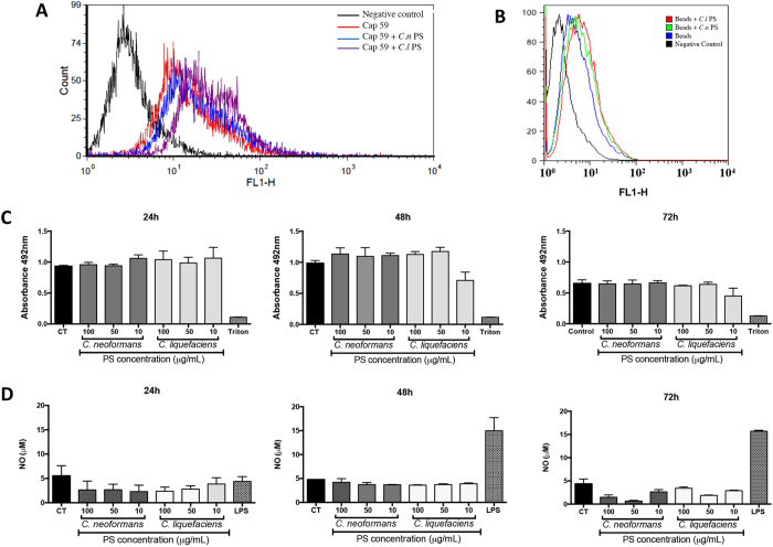
Figure 3. Phagocytosis by murine macrophages of acapsular Cryptococcus neoformans cells (cap59 mutant) or beads coated with secreted PS.
(A,B) Macrophages were allowed to interact, for 72 h, with FITC-labelled Cap59 (A) or polystyrene beads (B) (coated with secreted-PS from C. neoformans (Cn-PS) or C. liquefaciens (Cl-PS), and then analyzed by flow cytometry. Non-infected macrophages were used as controls (CT). (C) Cell viability (by the XTT assay) and nitric oxide production (D) of murine macrophages exposed to secreted-PS from C. neoformans and C. liquefaciens for 24, 48 and 72 h. Treatment with Triton X-100 and LPS were used as positive controls for cell viability loss and macrophage activation, respectively. In (C and D) data represent mean ± SD of 3 independent experiments.

