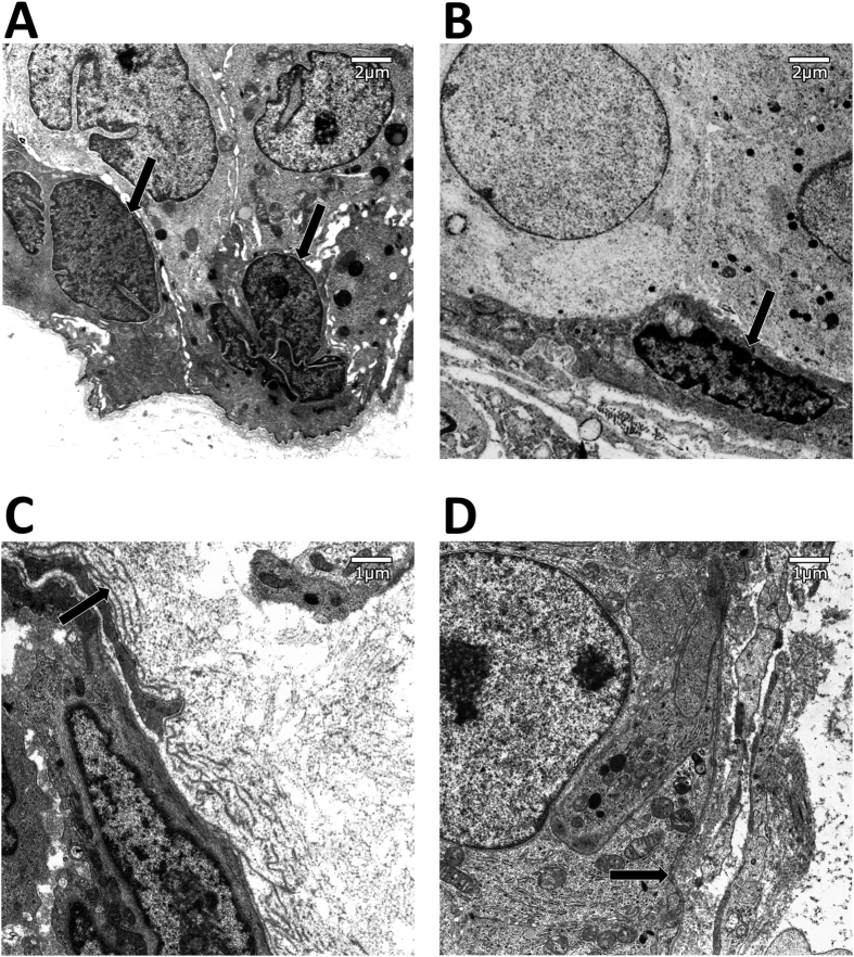
Figure 5. Electron micrograph of myoepithelial cell nucleus.
Myoepithelial cells in DCIS lesions have a flatter nucleus than in UDH. Arrow: Myoepithelial cell nucleus. (A) Myoepithelial cell nucleus in a benign case. (B) Myoepithelial cell nucleus in a DCIS case. Electron micrograph of basal lamina. In benign ducts, the basal lamina has multiple layers, while in DCIS it has a single layer. Arrow: Basal lamina. (C) Basal lamina in a benign lesion. (D) Basal lamina in a DCIS lesion.

