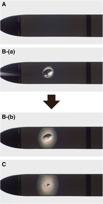
Fig. 2.

Embryo vitrification procedures using the Kitasato Vitrification System. A: The vitrification solution absorber of the device is set on a stereomicroscope. B: After an embryo with a small volume (≤0.4 μL) of vitrification solution is placed dropwise on the vitrification absorber (a) that embryo can easily be observed under the stereomicroscope (b). C: Subsequently, the vitrification solution surrounding the embryo is absorbed by the absorber within less than 5–6 s, and the embryo, with a small remaining volume of vitrification solution, can easily be observed
