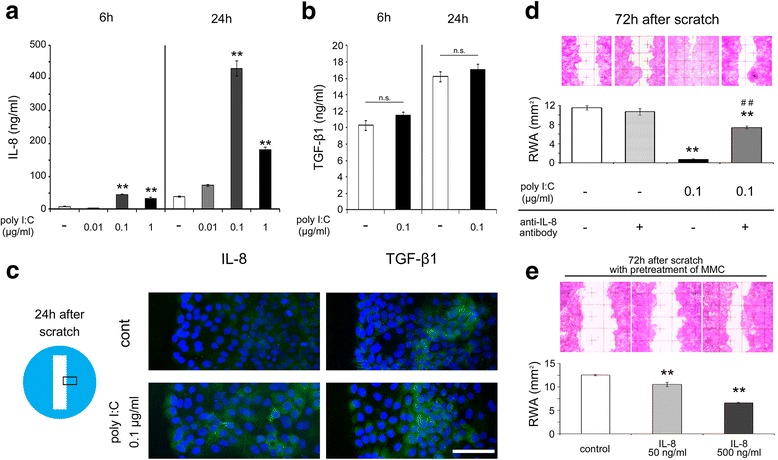
Fig. 3.

The protein levels and immunohistochemistry results of IL-8 and TGF-β1 and the involvement of IL-8 in migration. a The IL-8 protein concentrations after 6 or 24 h of incubation in the culture medium following the scratch and treatment with 0.1 μg/ml of poly I:C (n = 3). b The TGF-β1 protein concentrations after 6 or 24 h of incubation in the culture medium following the scratch and treatment with 0.1 μg/ml of poly I:C (n = 3). c Representative immunoreactivity of IL-8 (green) or TGF-β1 (green) on the scratched edge margins with or without 0.1 μg/ml of poly I:C stimulation at 24 h after the scratch (Blue; DAPI). d Representative images showing the HaCaT cell remaining wound area at 72 h after the scratch with anti-IL-8 antibody and the remaining wound area in a 3 × 8 mm2 rectangle (n = 3). e Representative images showing the HaCaT cell migration at 72 h after the scratch with human recombinant IL-8 and the remaining wound area in a 3 × 8 mm2 rectangle (n = 3). Data are presented as the mean ± SEM. * p < 0.05 vs. control, ** p < 0.01 vs. control, ## p < 0.01 vs. poly I:C alone according to the Tukey-Kramer test (a, d) or the Mann–Whitney test (b). Data for the immunofluorescence assays are representative of at least three independent experiments. Grids = 1 × 1 mm2. Scale bar: 100 μm. RWA; remaining wound area. MMC; mitomycin C
