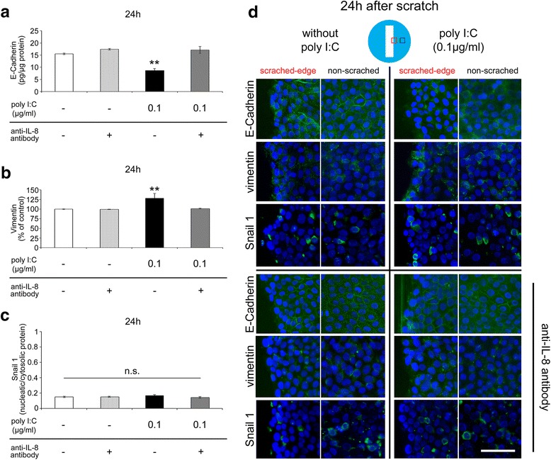
Fig. 4.

EMT-associated cellular marker alterations. a E-Cadherin (epithelial cell marker) protein levels at 24 h after scratching (n = 4). b Vimentin (mesenchymal cell marker) protein levels at 24 h after scratching (n = 4). c The nucleus/cytosol ratio of Snail 1 (EMT-related transcriptional factor) protein levels at 24 h after scratching (n = 4). d Immunoreactivities of E-cadherin (green), vimentin (green), and Snail 1 (green) at 24 h after the scratch (Blue; DAPI) 24 h after poly I:C stimulation with or without anti-IL-8 antibody. Data are presented as the mean ± SEM. n.s.: not significant according to the Tukey-Kramer test. Data for immunofluorescence assay are representative of at least three independent experiments. Scale bar: 100 μm
