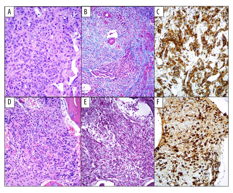
Figure 2.
(A) H&E, 400×. Liver biopsy showing extensive involvement by acute megakaryoblastic leukemia. The liver architecture is distorted by the presence of numerous aggregates of large, markedly atypical neoplastic cells. (B) Trichrome special stain, 200×. Extensive collagen deposition. (C) CD42b, 400×. The atypical megakaryocytes are highlighted with immunohistochemical stains for CD42b. (D) H&E 400×. Cellular bone marrow with small hypolobated or immature megakaryoblasts. (E) Reticulin special stain, 400×. Marked bone marrow fibrosis. (F) CD42b, 400×. The atypical megakaryocytes are highlighted with immunohistochemical stains for CD42b.

