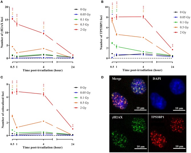
Figure 3.
Irradiation induces endothelial DNA damage signaling. Graphs represent the amount of γH2AX (A), TP53BP1 (B) and colocalized γH2AX/TP53BP1 (C) foci 30 min, 1, 4, and 24 h after irradiation with a single X-ray dose of 0.05, 0.1, 0.5 and 2 Gy (n = 6). Data show means ± SEM. *P < 0.05, †P < 0.005, ‡P < 0.001 compared to sham on the same day, using 2-way ANOVA with Bonferroni post-hoc test. (D) Representative images showing γH2AX (green), TP53BP1 (red) and γH2AX+TP53BP1 (yellow) foci in DAPI stained nuclei (blue) of TICAE cells 30 min after irradiation with a single X-ray dose of 2 Gy.

