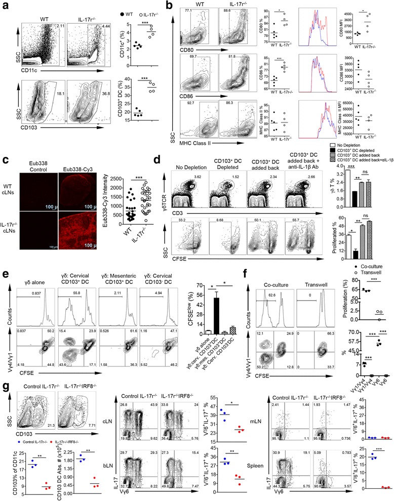
Fig. 4.

CD103+ DCs specifically induce Vγ6 γδT17 cell proliferation. a After ex vivo staining of cLN cells and gating from total live cells, CD11c+ cells were gated for calculating total DCs (top) then from this population CD103+ DCs were gated (bottom). Representative of two to three experiments. ***p < 0.001. b Gated on total CD103+ DCs from WT and IL-17r−/− cLNs, staining of surface activation markers CD80, CD86, and MHC Class II was performed. Both percentages and MFI are shown. Representative of two to three experiments. *p < 0.05, ***p < 0.001. c 16S rRNA FISH hybridization on frozen tissue sections from WT and IL-17r−/− cLNs. Probe Eub338-cy3 was used and TRIC signal intensity at random, yet the same locations for WT and IL-17r−/− cLN was used to quantify the amount of bacterial RNA in LNs. Representative of three different cLNs from three mice. ***p < 0.001. d CFSE-labeled whole cLN cells were depleted of CD103+ DCs and cultured for 5 days. In conditions of whole cLN cells depleted with CD103+ DCs, sorted CD103+ DCs were added back in the presence or absence of anti-IL-1β neutralizing mAb (2 μg/ml). γδT cell proliferation was shown. Cells were gated on 7AAD−CD3+γδTCR+ cells. Representative of five experiments. *p < 0.05, **p < 0.01, ***p < 0.001. n.s. not significant. e Using MoFlo sorter, WT γδ T cells from the LNs and spleens were purified and CFSE labeled then co-cultured with three different IL-17r−/− DC populations (CD103+ cLNs, CD103+ mLNs, and CD103− cLNs) for 5 days. Representative histograms of γδT cell proliferation and summarized percentages of proliferated cells are shown (upper panel). Representative dot plots of proliferated γδT cells with Vγ4/Vγ1 staining are shown (bottom panel). Cells were gated on 7AAD−CD3+γδTCR+ cells. Representative of three experiments. *p < 0.05. f 96-well transwell plate was utilized to separate CD103+ DCs from cLNs of IL-17r−/− mice from WT γδ T cells to determine if cell-to-cell contact is required. Representative histograms of γδT cell proliferation and summarized percentages of proliferated cells are shown (upper panel). Representative dot plots of proliferated γδT cells with Vγ4/Vγ1 staining and summarized data are shown (bottom panel). Cells were gated on 7AAD−CD3+γδTCR+ cells. Representative of three experiments. ***p < 0.001. g After immunostaining of cLNs, gated from total CD11c+ DCs then gated on CD103+ DC population to analyze total CD103+ DC % and absolute number. After PMA/ionomycin stimulation and intracellular staining, gated from total γδ T cells then gated on Vγ6+ and IL-17+ to calculate total Vγ6+ γδT17 %. Representative of two experiments. *p < 0.05, **p < 0.01, ***p < 0.001
