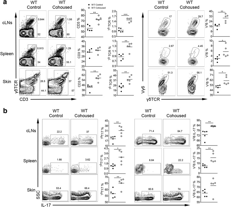
Fig. 6.

Co-housing of IL-17r−/− mice with WT mice induces the proliferation of γδT17 cells in the cLNs and systemic expansion of γδT17 cells in WT mice. a Flow cytometry analysis of cell homogenate from different tissues examining the surface marker percentages of CD3, γδ T cells and Vγ6 γδ T cells between control and co-housed WT mice with IL-17r−/− mice of the same age and sex. Representative dot plots and summarized data showing percentages of total CD3+ T cells, γδT cells, and Vγ6 T cells. *p < 0.05, **p < 0.01, ***p < 0.001. b Intracellular IL-17 expression between control and co-housed WT mice with IL-17r−/− mice of the same age and sex. Representative dot plots and summarized data are shown. *p < 0.05, **p < 0.01
