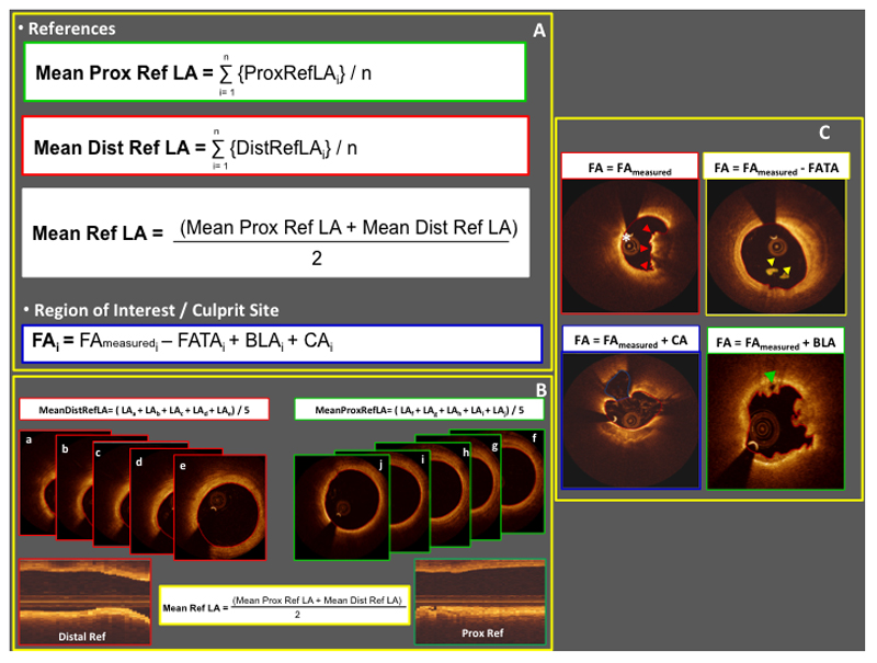
Figure 2. Pre-stenting FD-OCT run measures.
Panel A and B. Mathematical formulae adopted to calculate mean reference lumen areas and flow areas. Panel C. Calculation of flow area according to the presence of thrombus adherent to the vessel wall (red arrowheads), floating thrombus (yellow arrowheads), cavity associated with plaque rupture (white asterisk), and bridging lumen within the thrombus (green arrowhead). Note that contours of meausured flow area are highlighted in red in each image of Panel C. (BLA: bridging lumen area; CA: cavity area; FA: flow area; FAmeasured: measured flow area; FATA: floating atherothrombus area; LA: lumen area; ref: reference; ROI: region of interest)

