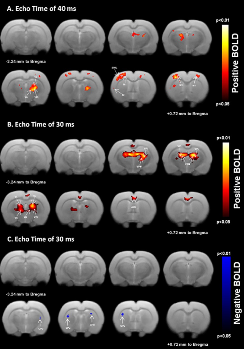
Fig 4. BOLD responses to unilateral sensorimotor electrical hindpaw stimulation.

Statistical maps calculated from group analysis (n = 12; 0.05≤p-value≤0.01) are overlaid onto a template rat brain (eight axial slices centered at -3.24 mm to Bregma to +0.72 mm to Bregma). Unilateral stimulation resulted in positive (A and B) and negative (C) BOLD responses in cortical and subcortical regions relevant for sensorimotor processing. Positive activated areas include the contralateral primary/secondary hindpaw somatosensory (S1HL/S2), bilateral primary motor (M1) cortex (A) as well as the bilateral sensorimotor thalamic nuclei (VPL and VA/VL) at TE = 40ms (A), larger positive activations are localized in the bilateral sensorimotor thalamic nuclei (VPL and VA/VL) at TE = 30ms (B). Negative activations are bilaterally measured in the dorsolateral caudate-putamen (CPu) region at TE = 30ms (C).
