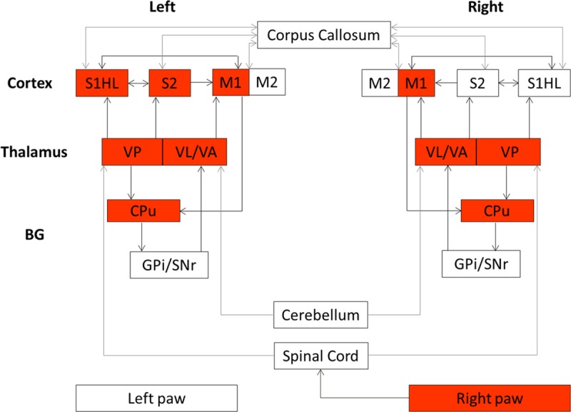
Fig 5. The rat brain sensorimotor network.

A simplified description derived from previous studies [1–7]. Both brain hemispheres are presented and arrows represent connections. Cortical regions include the hindpaw primary/secondary somatosensory cortex (S1HL/S2) and the primary /secondary motor cortex (M1/M2). Subcortical regions include the thalamus represented by the posteroventral thalamic nucleus (VP), the motor thalamic nucleus including the ventral lateral thalamic nucleus (VL) and the ventral anterior thalamic nucleus (VA) and Basal Ganglia (BG) represented by the caudate putamen (CPu), the substantia nigra-pars reticulata (SNr) and the internal segment of the globus pallidus (GPi). Red boxes highlight the elicited structures after unilateral sensorimotor hindpaw stimulation in the present study.
