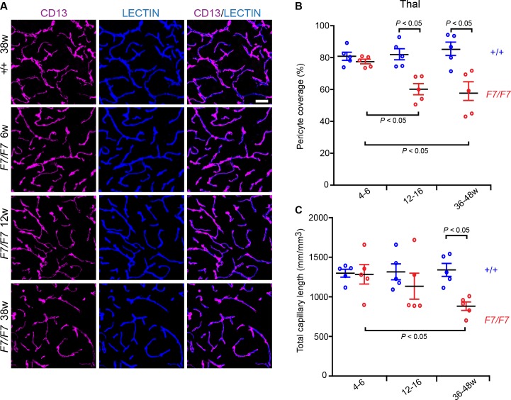
Fig 3. Loss of pericyte coverage and capillary reductions in the posterior thalamus of adult PdgfrβF7/F7 (F7/F7) mice.
(A) Representative confocal microscopy analysis of coronal sections showing CD13-positive pericyte coverage (magenta, left panels), lectin-positive endothelial vascular profiles (blue, middle panels) and merged (right panels) in the posterior thalamus of 6-, 12- and 36-week old F7/F7 and 36-week old control mice (+/+ 36w). Bar = 40 μm. (B-C) Quantification of pericyte coverage (B) and total capillary length (C) in 4–6, 12–16, and 36-48-week old F7/F7 mice compared to age-matched littermate controls (+/+) determined as in Fig 1. Mean ± S.E.M., n = 5 animals per group. In B and C, one-way ANOVA and Bonferroni’s post hoc tests were used to compare data in F7/F7 mutants versus age-matched littermate controls, and/or between different age groups of F7/F7 mutants only. P < 0.05 indicates statistically significant differences between groups.

