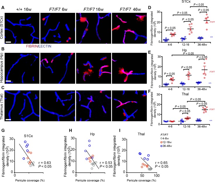
Fig 6. Accumulation of fibrinogen and fibrin perivascular deposits in the brain of adult PdgfrβF7/F7 (F7/F7) mice.
(A-C) Representative confocal images of lectin-positive endothelial profiles and extravascular fibrinogen and fibrin deposits in the somatosensory cortex S1 region (S1Cx) layer IV (A), the CA1 region stratum pyrmidale of the hippocampus (Hp) (B) and posterior thalamus (C) of 6-, 16- and 46-week old F7/F7 mice and 16-week old control (+/+) mice. Bar = 20 μm. (D-F) Quantification of fibrinogen and fibrin-positive extravascular deposits in the S1Cx (D), Hp (E) and Thal (F) of 4–6, 12–16, and 36-48-week old F7/F7 mice and age-matched littermate controls (+/+). Mean ± SEM, n = 5 mice per group. In each animal, 4–6 randomly selected fields in the cortex, hippocampus and thalamus were analyzed in 4 non-adjacent sections (~100 μm apart) and averaged to calculate individual values per mouse. In D-F, one-way ANOVA and Bonferroni’s post hoc tests were used to compare data in mutants versus age-matched littermate controls, and/or between different age groups of F7/F7 mutants. P < 0.05 indicates statistically significant differences between groups. (G-I) Correlations between age-dependent fibrinogen and fibrin perivascular accumulation and loss of pericyte coverage in the S1Cx (G), Hp (H), and Thal (I) regions in F7/F7 mice. Single data points were from 4-6- (grey), 12-16- (red), and 36-48- (blue) week old F7/F7 mice (n = 15 individual points per mouse; r = Pearson’s coefficient; P, significance.

