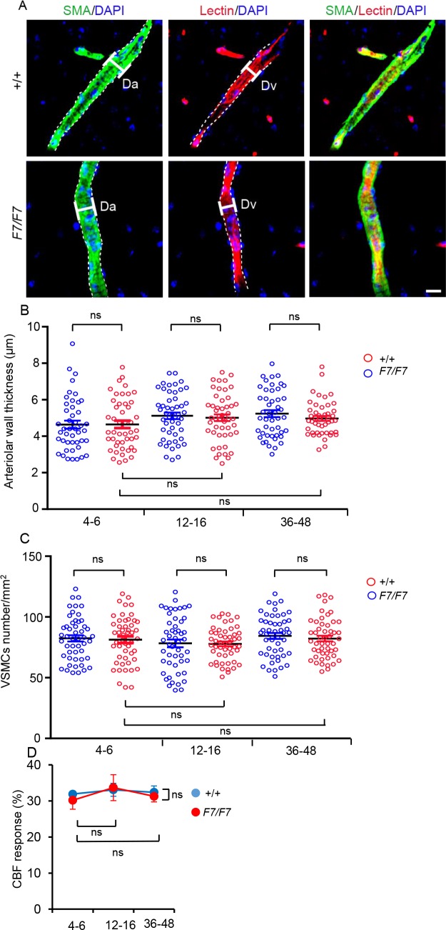
Fig 7. Normal thickness of vascular smooth muscle cells (VSMCs)-covered arteriolar wall, VSMCs cell numbers and functional response to adenosine in adult PdgfrβF7/F7 (F7/F7) mice.
(A) Representative high magnification confocal images (see Methods for details) of α-smooth muscle actin (SMA)-positive VSMCs (green, left panels) and lectin-positive endothelial profiles (red, middle panels), and merged (left panels) in penetrating arterioles of the somatosensory cortex S1 layer 1 cortex (S1Cx) of 12-week old F7/F7 and age-matched littermate control mice (+/+). Interrupted lines indicate the edge of fluorescent signal. Da, SMA-positive arteriolar diameter; Dv, lectin-positive endothelial diameter. Scale bar = 50 μm. (B-C) Quantification of the arteriolar wall thickness (B) and VSMCs numbers (C) in 4-6-, 12-16- and 36-48-week old F7/F7 mice compared to the respective age-matched littermate controls (+/+). In B and C, individual points represent 10 vessels per animal from 5 animals per group. Mean ± S.E.M. from 50 arterioles per group. ns, not significant by one-way ANOVA. See Methods for details. (D) Laser Doppler flowmetry measurements of cerebral blood flow (CBF) response to endothelium-independent VSMCs relaxant adenosine (400 μM) in 4-6-, 12-16-, and 36-48-week old F7/F7 mice compared to age-matched littermate controls (+/+). Mean ± S.E.M.; n = 3 mice per group; ns, not significant by one-way ANOVA.

