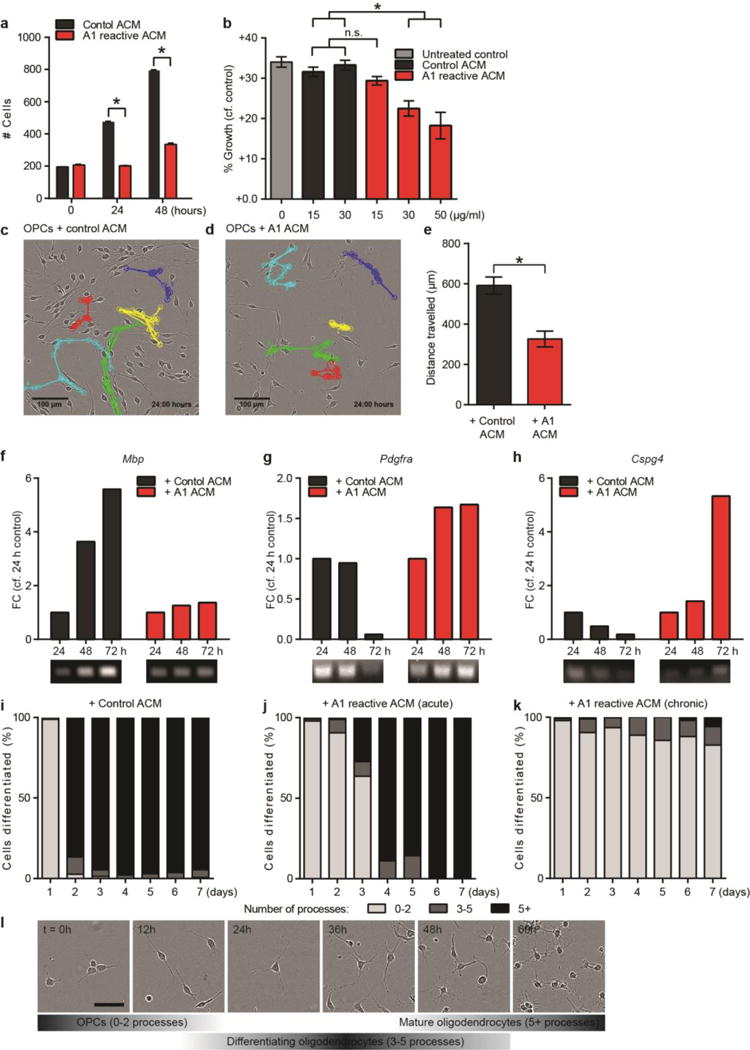
EXTENDED DATA FIGURE 10. A1 reactive astrocytes inhibit oligodendrocyte precursor cell proliferation, differentiation and migration.

a, Number of cells counted from phase-contrasted images of oligodendrocyte precursor cells (OPCs) treated with control and A1 reactive conditioned media (ACM). b, EdU ClickIt® assay determined growth of OPCs treated with increasing concentration of control and A1 ACM for 7 days. Both a and b show A1 ACM decreases OPC proliferation compared to control. c, d, Representative images of tracked OPC migration following treatment with control (c) and A1 (d) ACM, quantified in e. N = 100 cells from 10 separate experiments. f–h, Representative RT-PCR ethidium bromide gel showing no increase in mature OL marker Mbp transcript in OPCs treated with A1 ACM, with no change in OPC marker Pdgfra and Cspg4 expression – evidence of a lack of differentiation into mature oligodendrocytes. Treatment of OPCs with control ACM did not delay their differentiation into mature oligodendrocytes. N = 2. i–k, Total number of terminal process of oligodendrocyte lineage cells were counted as a measure of differentiation. Over 90% of cells differentiated by 24 h after removal of PDGFα when treated with control ACM (i). In contrast, treatment with a single dose (j) or daily doses (k) of A1 ACM delayed this level of differentiation by 72 h following a single dose, or indefinitely with chronic treatment. N = 6 separate experiments. l, representative phase images and time scale for oligodendrocyte differentiation assay (treated with control ACM). Scale bar: 100 μm (c,d), 25 μm (l). * p < 0.05, one-way ANOVA, except panel e (Student’s t-test). Error bars indicate s.e.m.
