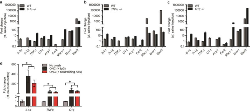
EXTENDED DATA FIGURE 11. Activation of microglia following lipopolysaccharide injection in knockout mice.

Mice from single global knock-outs of Il-1α (a), TNFα (b), and C1q (c) were treated with lipopolysaccharide (5 mg/kg, i.p.) and microglia collected 24 h later. Single knock-out animals still showed upregulation of many markers of microglial activation, as determined by qPCR. N = 3 for Il-1α and C1q, N = 5 for TNFα. d, quantitative PCR for microglia-derived A1-inducing molecules in the optic nerve of mice that received an optic nerve crush. Following crush, optic nerve contained neuroinflammatory microglia, while injection of A1 astrocyte-neutralizing antibodies into the vitreous of the eye did not decrease microglial activation (however it did halt A1 astrocyte activation in the retina – see Fig. 4). Error bars indicate s.e.m. * p < 0.05, one-way ANOVA.
