
Figure 2. A1 reactive astrocytes do not promote synapse formation or function.
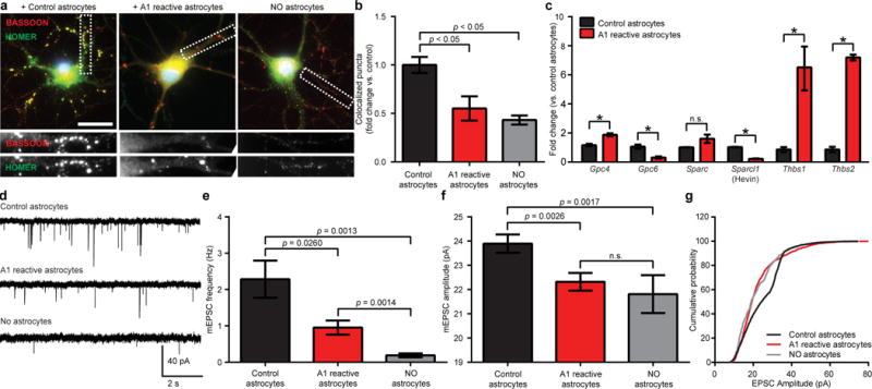
a, Representative images of retinal ganglion cells (RGCs) grown without astrocytes, or with control or A1 reactive astrocytes, immunostained with pre- and post-synaptic markers HOMER (green) and BASSOON (red). Co-localization (yellow puncta) was counted as a structural synapse. b, Total number of synapses normalized per each individual RGC, n = 50 neurons in each treatment. c, Quantitative PCR for astrocyte secreted synaptogenic factors. d, Representative traces of whole-cell patch clamp mEPSC recordings from RGCs. e, Frequency of mEPSCs was significantly decreased in presence of A1s (RGCs without astrocytes: 0.19 ± 0.05 Hz n = 12 neurons, RGCs with resting astrocytes: 2.28 ± 0.51 Hz n = 14 neurons, RGCs with A1s: 0.95 ± 0.19Hz n = 16 neurons). f, A1s significantly decreased mean amplitude of mEPSCs (RGCs without astrocytes: 21.81 ± 0.78 pA n = 12 neurons, RGCs with resting astrocytes: 23.89 ± 0.38 pA n = 14 neurons, RGCs with A1s: 22.32 ± 0.37 pA n = 16 neurons). g, RGCs cultured with A1s had significantly more small amplitude mEPSCs in cumulative probability histograms (p < 0.0001 Kolmogorov-Smirnov test, n = 12–16 neurons per condition). * p < 0.05, one-way ANOVA. Error bars indicate s.e.m. Scale bar: 10 μm.