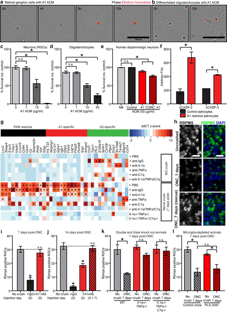
Figure 4. Astrocyte-derived toxic factor promoting cell death.

Representative phase image showing death of purified retinal ganglion cells (RGCs) in culture (ethidium homodimer stain in red shows DNA in dead cells (a, 24 h quantification in c), and differentiated oligodendrocytes (b, 24 h quantification in d). e, Quantification of A1-induced cell death in human dopaminergic neurons (5 days). f, Western blot analysis for cleaved caspase-2 and -3 in RGCs treated with control and A1 ACM. g, retro-orbital optic nerve crushes (ONC) produced A1s in the retina. Intravitreal injection of neutralizing antibodies to Il-1α, TNFα, and C1q blocked A1 production. h, RBPMS (RNA-binding protein with multiple splicing, an RGC marker) immunostaining of whole-mount retinas showed decreased number of RGCs in ONC, rescued with neutralizing antibody treatment. Quantification is shown post ONC at 7 days (i), 14 days (j) using neutralizing antibodies, and at 7 days using Il1α−/−TNFα−/− and Il1α−/−TNFα−/− C1q−/− animals (k), and microglia-depleted (PLX-3397-treated) animals (l). * p < 0.05, one-way ANOVA. n = 8 in each instance. Error bars indicate s.e.m. Scale bar: 100 μm (a,b); 20 μm (k).
