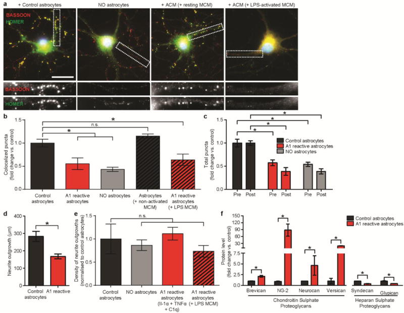
EXTENDED DATA FIGURE 6. A1 reactive astrocytes do not promote synapse formation or neurite outgrowth.

a, Representative images of retinal ganglion cells (RGCs) grown without astrocytes, or with control or A1 reactive astrocytes, stained with pre- and post-synaptic markers HOMER (green) and BASSOON (yellow). Colocalization of these markers (yellow puncta) was counted as a structural synapse. b, Total number of synapses normalized per each individual RGC. The number of synapses decreased after growth of RGCs with LPS-activated microglial conditioned media (MCM)-activated A1 reactive astrocyte conditioned media (ACM), or Il-1α, TNFα, C1q-activated A1 reactive astrocytes was not different. N = 50 neurons in each treatment. c, Quantification of individual pre- and post-synaptic puncta. d, Total length of neurite growth from RGCs. e, Density of RGC processes in cultures used in measurement of synapse number. There was no difference in neurite density close to RGC cell bodies (where synapse number measurements were made). f, Western blot analysis of proteoglycans secreted by control and A1 reactive astrocytes. Conditioned media from control astrocytes contained less chondroitin sulphate proteoglycans Brevican, Ng2, Neurocan and Versican, while simultaneously having higher levels of heparan sulphate proteoglycans Syndecan and Glypican. * p < 0.05, one-way ANOVA, except d (Student’s t-test). Scale bar: 10 μm. Error bars indicate s.e.m.
