
Figure 3. C1 neuron stimulation protects against renal IRI.
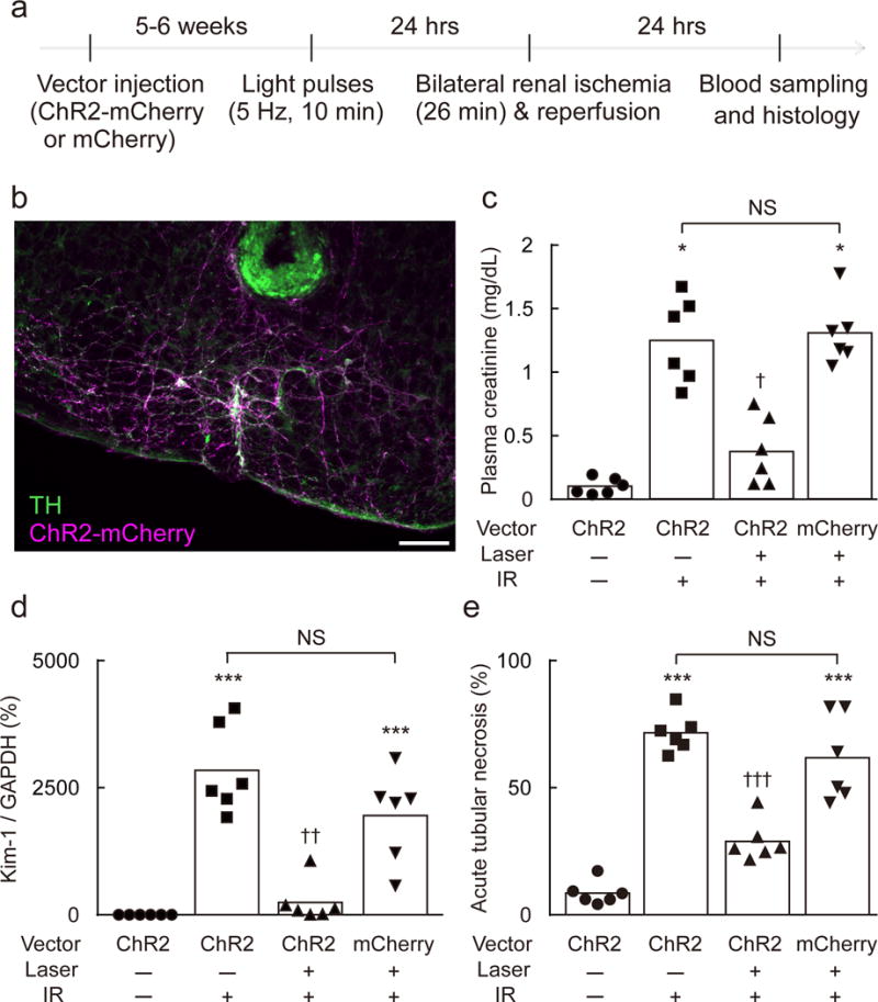
(a) Time line of experiment. (b) Section through the rostral medulla oblongata of a DBH-cre mouse 5–6 weeks after local injection of AAV2–DIO–EF1α–ChR2–mCherry. mCherry (magenta) and tyrosine-hydroxylase (TH, green) immunoreactivities are colocalized (white). Most catecholaminergic neurons contain mCherry. A small lesion surrounded by green autofluorescence shows the location of the tip of the optical fiber (scale bar: 100 μm). (c–e) Effect of prior C1 neuron stimulation on plasma creatinine, Kidney Injury Molecule-1 (Kim-1) mRNA (Havcr1/Gapdh ratio) in the kidney, and acute tubular necrosis (% of kidney section surface area) in DBH-cre mice after IRI (n = 6/group). Vector used: AAV2–DIO–EF1α–ChR2–mCherry (ChR2) or AAV2–DIO–EF1α–mCherry (mCherry). Laser: 5 Hz, 10 min. IR, ischemia-reperfusion. Statistical analysis (Kruskal-Wallis with Steel-Dwass test): [H = 18.62, P = 0.0003] (c), [H = 19.22, P = 0.0004] (d), [H = 19.77, P < 0.0001] (e), * vs. ChR2:Laser(−):IR(−); † vs. ChR2:Laser(−):IR(+) or mCherry:Laser(+):IR(+). Single, double, or triple significant symbols indicate P < 0.05, P < 0.01, or P < 0.001, respectively.