
Figure 4. C1 neurons mediate the protective effect of restraint stress against renal ischemia-reperfusion injury (IRI).
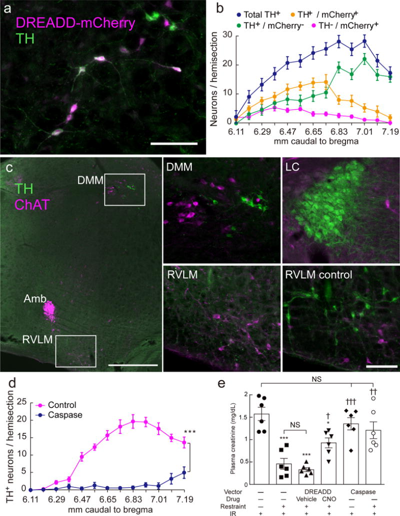
(a) mCherry (magenta) and TH (green) immunoreactivity in the left medulla oblongata of a DBH-cre mouse six weeks after stereotaxic microinjection of AAV2–DIO–hSyn–hm4D(Gi)–mCherry (DREADD) (transverse section; scale bar: 100 μm). (b) Rostrocaudal distribution (mm caudal to bregma) of mCherry and TH immunoreactivities (n = 12). (c) Choline acetyltransferase- (ChAT; magenta) and TH- (green) immunoreactivities in the left medulla oblongata of a DBH-cre mouse 6 weeks after injection of AAV2–DIO–taCasp3–TEVp (AAV2-caspase). After AAV2-caspase treatment, the C1 neurons in the rostral ventrolateral medulla (RVLM) are undetectable (left and lower middle panels) but other catecholaminergic neurons (dorsal medulla, DMM and locus coeruleus, LC) are intact (top middle and top right panels). ChAT+ neurons are unaffected by AAV2-caspase treatment regardless of location (left and bottom middle). Scale bar: 500 μm (left) or 100 μm (four right panels). Amb, nucleus ambiguus. (d) Rostrocaudal distribution of TH-immunoreactive RVLM neurons in control DBH-cre mice (n = 7) vs. caspase-treated DBH-cre mice (n = 10). Lesions were bilateral; cells were counted on one side only. Statistics: two-way ANOVA with Tukey–Kramer test; [F(12, 180) = 25.99, P < 0.0001]. (e) The protective effect of restraint stress against renal IRI was attenuated by inhibiting (DREADD) or lesioning (caspase) the C1 neurons (n = 6 DBH-cre mice/group). CNO: clozapine N-oxide (3 mg/kg); Vehicle: saline. Statistics: one-way ANOVA with Tukey–Kramer test; [F(5, 30) = 14.11, P < 0.0001]. * vs. Restraint(-):IR(+) and † vs. Restraint(+):IR(+) and DREADD:Vehicle:Restraint(+):IR(+). Single, double, or triple significant symbols indicate P < 0.05, P < 0.01, or P < 0.001, respectively.