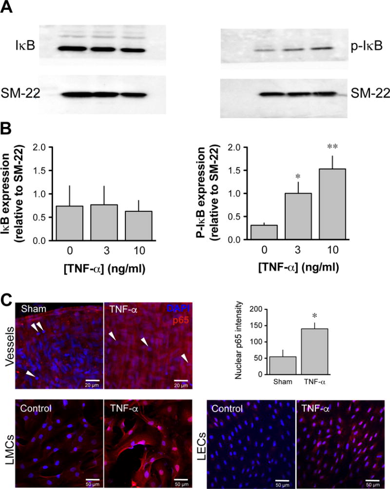
Figure 3. Effect of TNF-α on NF-κB activation in lymphatic vessels.

A. Representative western blots of unphosphorylated (left panel) and phosphorylated (right panel) IκB protein following 4-h incubation without (sham) and with 3ng/ml and 10 ng/ml TNF-α. B. Summary densitometry bar graph of the level of IκB and p-IκB in the three vessel groups normalized to the level of SM22 (n=4; *P<0.05, **P<0.01, repeated measures of ANOVA with Dunnett’s post-hoc test). C. Level of nuclear translocation of the NF-κB subunit, p65 in lymphatic cells, illustrated by confocal immunofluorescence. Representative images depicting nucleus (blue, DAPI) and p65 (red) in a sham (top left panel) and a TNF-α-treated lymphatic vessel (middle panel, 10 ng/ml, 3-h incubation). Maximal projection of a stack of 3 images taken at the level of the muscles, as denoted by the vertical (perpendicular to the vessel length) orientation of most of the nucleus (note immune cells identified by their round nucleus, arrowheads). Nuclear translocation (pink nuclear staining) is apparent in most cells of the TNF-α-treated vessel as confirmed by the summary bar graph representing the level of nuclear p65 fluorescence (right panel; n=4; *P<0.05 vs own sham, paired Student t-test). Nuclear translocation of the p65 subunit is also evident in cultures of lymphatic endothelial (bottom left panels) and muscle cells (bottom right panels) isolated from the same rat mesenteric vessels when treated with TNF-α. Images are representative of 3–5 similar experiments.
