Fig 4.
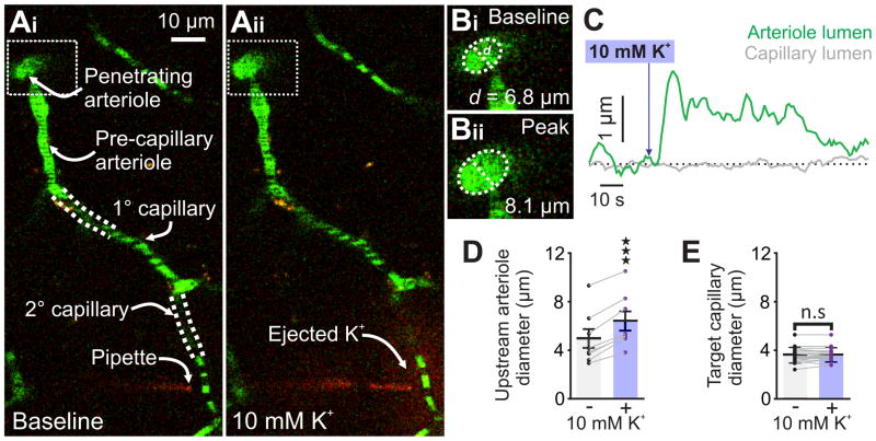
10 mM K+ applied to capillaries causes upstream arteriolar dilation in vivo. (Ai) Micrograph illustrating pipette placement adjacent to a second-order capillary for in vivo monitoring of the diameter of the upstream feed arteriole (boxed). White dashed lines indicate that capillaries are connected into a single tree despite deviating from the imaging plane. (Aii) The same experiment, after pressure ejection of 10 mM K+ and TRITC (red) around the capillary. Note the dilation in the feed arteriole (boxed). (B) Magnification of the boxed areas around the feed arteriole in Ai and Aii, illustrating the magnitude of dilation evoked by capillary stimulation with 10 mM K+. (C) Traces illustrating the luminal diameter of a pre-capillary arteriole (green) and the stimulated capillary (grey) before and after stimulation with 10 mM K+. Delivery of K+ produced a robust dilation in the arteriole, but not the capillary. (D) Summary data showing arteriole diameter before and after capillary application of 10 mM K+, which produced significant upstream arteriole dilation (n = 8 paired experiments, 7 mice; ****P < 0.0001 (t7 = 10.86) paired Student’s t-test). (E) Summary data showing target capillary diameter before and after capillary application of 10 mM K+, which had no effect (n = 18 capillaries, 7 mice P = 0.6014 (t17 = 0.5324) paired Student’s t-test). All error bars represent s.e.m.

