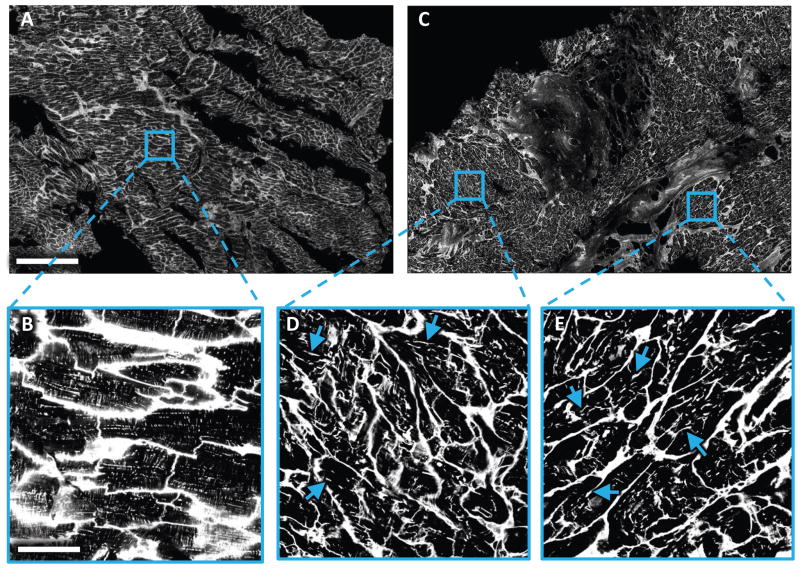
Figure 1. Two-dimensional tile scan of WGA-labeled LV tissue slices obtained from control and HF patient at time of LVAD implantation.
Images were acquired with a confocal microscope equipped with a 40× oil immersion lens using a pixel size of 0.2×0.2μm. (A) Tile scan from donor tissue. (B) Magnified view of the boxed region in (A) showing myocytes with dense t-system. (C) Tile scan from HF tissue. (D,E) Magnified views of the boxed region in (C) with myocytes with a sparse and irregular t-system. The remodeled t-system exhibited longitudinal components in the majority of cells. Some examples were marked with arrows. Scale bar in A is 500μm and also applies to C. Scale bar in B is 50μm and also applies to D and E.

