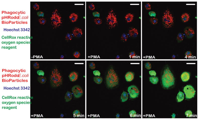
Figure 3. hiPSC-microglia demonstrate functional abilities consistent with human microglia.
Confocal imaging of hiPSC-MG reveals a noticeable increase in phagocytosis of pHrodo™ Red E. coli BioParticles (red fluorescence) without treatment with phorbol myristrate acetate (-PMA), however upon stimulation with PMA, they show production of reactive oxygen species (green fluorescence) starting at t=5 min. Hoechst 3342 (blue fluorescence) was used to visualize nuclei. The figure is representative of three independent experiments carried out in replicates (n=6). Scale bars, 50 μm.

