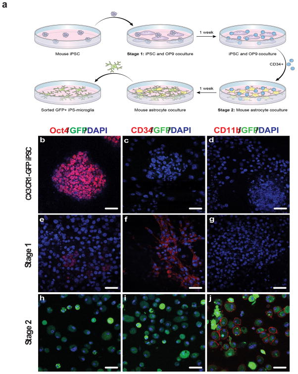
Figure 4. Mouse iPSCs are differentiated into microglia using a two-stage process.
(a) Schematic of the murine iPSC differentiation process to microglia-like cells. (b–j) Cx3cr1gfp/+ miPSCs, Stage 1 hematopoietic progenitor-like cells, and Stage 2 miPS-MG immunostained with antibodies to Oct4 (b,e,h), CD34 (c,f,i), CD11b (d,g,j), and endogeneous CX3CR1-GFP expression (b–j). DAPI was used to visualize nuclei. Cells expressing the CX3CR1-GFP reporter (h,i,j) only became evident at the completion of Stage 2 culture. The figure is representative of three independent experiments done in replicates (n=6). Scale bars, 50 μm.

