
Figure 5. Over-expressed and endogenous synuclein localizes to secretory granules in adrenal chromaffin cells.
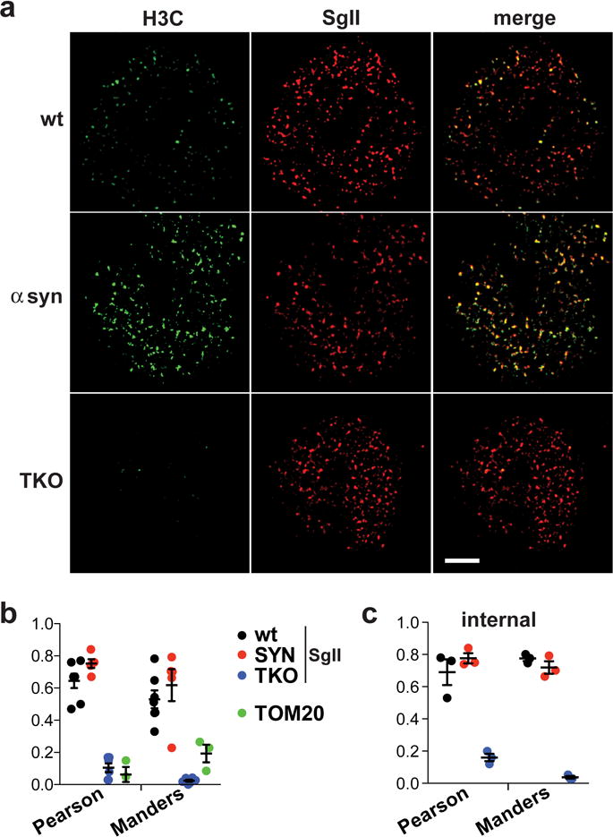
(a) Chromaffin cells from wt or synuclein TKO mice were transduced with lentivirus encoding either human α-synuclein (SYN) or empty vector, cultured for 72 h and immunostained for α-synuclein (H3C, green) as well as the dense core vesicle protein secretogranin II (SgII, red) The images were obtained using structured illumination and shown here as reconstructions of a 120 nm-thick slice located within 0.5 μm of the cell-coverglass interface. Size bar, 2.5 μm. (b) The extent of SgII colocalization with synuclein was quantified using Pearson’s correlation coefficient (R) and Manders overlap coefficient (M1). The extent of wt synuclein colocalization with the mitochondrial protein TOM20 is shown in green. n = 7 cells for wt, 5 cells for SYN, 6 cells for TKO and 3 cells for TOM20 (c) Similar colocalization measures for a slice located 0.5–1.0 μm deeper inside the cell shows that the localization of synuclein to secretory vesicles is not limited to the docked pool; n = 3 cells. Values in b and c indicate mean ± SEM