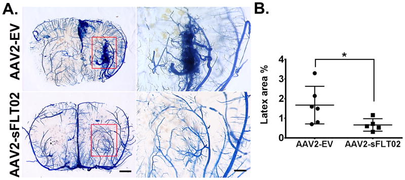
Figure 1. AAV2-sFLT02 in situ inhibits bAVM in Model 1 (R26CreER;Eng2f/2f).
(A) Representative images: coronal sections - latex-perfused brain. AVM developed in angiogenic regions (rectangles) of AAV2-EV-injected but not in AAV2-sFLT02-injected mice. Right panels: Close-up views: angiogenic region. Scale bars: 1 mm (left) or 500 μm (right). (B) Latex-perfused area quantification. AAV2-EV group: N=6. AAV2-sFLT02 group: N=5. *: p=0.037.

