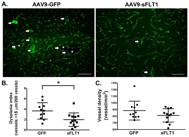
Figure 2. AAV9-sFLT1 IV reduces DI in bAVM in Model 1 (R26CreER;Eng2f/2f).
(A) Representative CD31 antibody-stained brain images. White arrows: Abnormal vessels. White stars: Enlarged abnormal vessel lumens. Scale bar=100 μm. (C) DI quantifications. (D) Vessel density quantifications. GFP-treated group: N=11. sFLT1-treated group: N=12. *: p=0.011.

