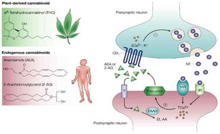
Figure 2.
The Endogenous Cannabinoid System. This figure illustrates how the endocannabinoid system transmits a signal from one neuron to another. The left panel shows the molecular structures of the plant derived (THC) and natural (AEA, 2AG) cannabinoids that bind to the CB1 cannabinoid receptor. The right panel illustrates how the endocannabinoid system works, using a schematic representation of a synaptic junction, which is where signals are passed from one neuron (presynaptic) to another (postsynaptic). When the presynaptic neuron is activated, it releases neurotransmitters (NT) that bind to receptors on the postsynaptic cell. Depending on the NT released, these can be ionotropic receptors (iR) which allow charged particles to flow directly into a cell, or metabotropic receptors (mR), which initiate a cascade of intracellular event events. In either case, intracellular calcium (Ca) is released, which stimulates the synthesis of endocannabinoids (AEA or 2AG) from precursor lipids located within the cell membrane. These endocannabinoids travel backwards to the presynaptic neuron where they bind to the CB1 receptors. Through a series of intracellular events, the endocannabinoids attenuate the subsequent release of neurotransmitter from the presynaptic neuron. Enzymes that breakdown the endocannabinoids (e.g. FAAH) are also located within this synaptic junction, enabling the rapid termination of the endocannabinoid signal. THC binds to the same CB1 receptors, displacing the natural cannabinoids, and remaining active for longer durations.
The figure is reprinted by permission from Macmillan Publishers Ltd: Nature Reviews Cancer. Guzman, M. Cannabinoids: potential anticancer agents. Nature Reviews Cancer, 3(10), 745–755., Copyright 2003.

