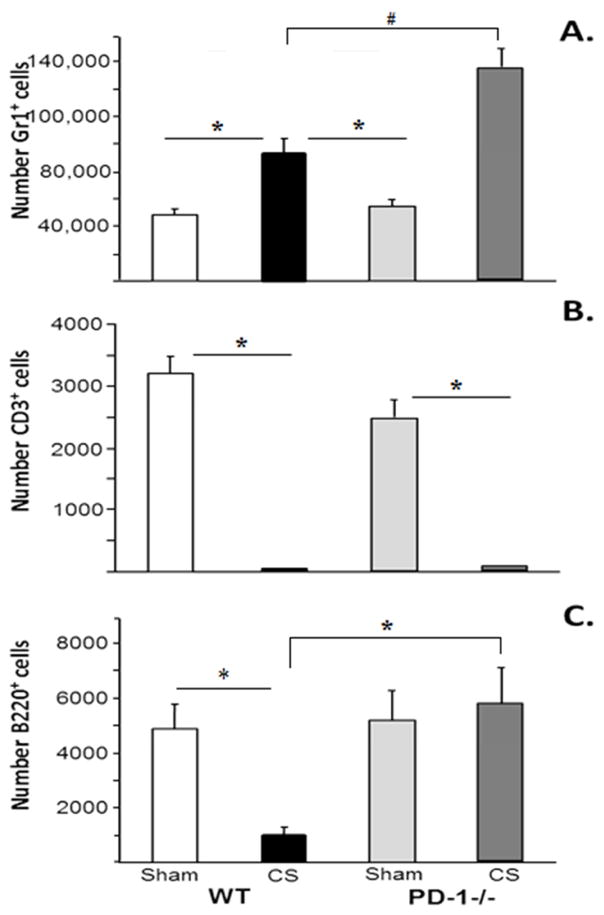
Figure 4a, 4b &4c.
Peritoneal cell influx in response to CS across WT and PD1−/− pups. 4a: Peritoneal neutrophil influx increased in WT, but was the neutrophil influx was more marked in PD-1−/− pups. 4b: Peritoneal lymphocyte loss was noted in both WT and PD-1−/− following CS. 4c: Peritoneal B-Cell population was decreased following CS in WT, but was preserved in PD-1−/− pups. N=6–9 per group. #p<0.05 comparing CS in PD-1−/− pups to all other groups. *=p<0.05; ANOVA.

