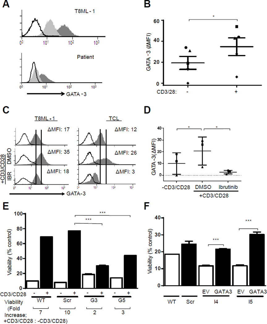
Figure 4.
TCR-mediated chemotherapy resistance is GATA-3 dependent. (A, B) T8ML-1 and primary TCL cells were activated with beads for 24 hours and GATA-3 expression in unstimulated (light gray histogram) and bead stimulated (dark gray histogram) cells determined by FACS analysis (isotype control, open histogram). Representative examples are shown in (A), and data obtained from 5 TCL patients summarized in (B). (C, D) TCL cells were activated in the presence of ibrutinib (1 µM) or vehicle control (DMSO) and GATA-3 expression determined in resting and bead stimulated cells. Representative examples are shown in (C) and data from 3 TCL patients summarized in (D). (E) T8ML-1 cells (wt) were transduced with either nontargeting (scr) or GATA-3 specific shRNA (G3, G5). Cells were cultured with or without beads, as indicated, in the presence of vincristine (6 nM). Cell viability was determined 48 hours later, and is reported relative to cells cultured without vincristine. The fold increase in viability between unstimulated and bead stimulated cells is shown below the x-axis. (F) GATA-3 was overexpressed in T8ML-1 transduced with ITK-targeting shRNA (I4, I5) following transduction with a vector containing full-length GATA-3 (GATA-3) or an empty vector (EV) control. Cells were cultured alone, or in the presence of vincristine (6 nM), and viability determined 48 hours later. Viability is reported relative to cells cultured without vincristine. (*p<0.05, **p<0.01, ***p<0.001 in unpaired two-sided Student t-test)

