FIG 6.
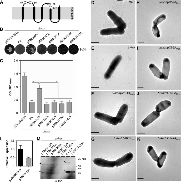
Functionality of M. tuberculosis VKOR in A. oris. (A) Presented is the membrane topology of M. tuberculosis VKOR. (B to K) The A. oris ΔVKOR mutant was transformed with plasmids expressing A. oris VKOR or M. tuberculosis VKOR or its Cys-to-Ala variants. The resulting strains were examined for their ability to aggregate with S. oralis (B), form biofilms (C), and assemble pili (D to K). Coaggregation and biofilm assays were performed as described in the legend to Fig. 2A and B. **, P ≤ 0.01. To examine pilus assembly, A. oris cells were subjected to EM as described in the legend to Fig. 3. Scale bars represent 0.5 μm. (L) The expression of the VKOR gene in A. oris ΔVKOR/pVKOR-2HA and A. oris ΔVKOR/pMtbVKOR was analyzed by quantitative RT-PCR. The results are presented as the average values from two independent experiments performed in triplicate. **, P ≤ 0.01. (M) Membrane fractions of the A. oris ΔVKOR mutant expressing M. tuberculosis VKOR or its variants, as well as A. oris VKOR-2HA or -C101A, were subjected to immunoblotting with anti-HA Ab in the absence of BME. Monomeric and complex forms of VKOR are marked by gray and black arrowheads, respectively.

