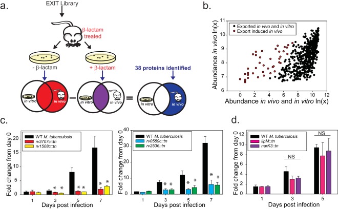
FIG 5 .
Strategy for identification of in vivo induced exported proteins. (a) Identification of in vivo induced exported proteins. Spleens from β-lactam-treated mice infected with the EXIT library were harvested after 2 weeks of infection. Spleen homogenates were plated in parallel on 7H10 agar without β-lactam to recover all clones (red Venn diagram) and on 7H10 agar containing β-lactam to recover clones exporting ‘BlaTEM fusion proteins during in vivo growth and in vitro growth (purple Venn diagram). The population of clones identified only or in significantly greater abundance on media lacking β-lactams represents proteins whose export was induced during infection (blue). (b) Sequenced read count values recovered from agar with or without β-lactam for the 593 EXIT proteins were plotted to compare abundances after β-lactam treatment in vivo, with the abundance after dual β-lactam treatment in vivo and in vitro indicated. The majority of proteins identified as exported in vivo remained highly abundant after additional β-lactam treatment in vitro (black). A total of 38 genes (highlighted in red) were identified as statistically less abundant after in vitro β-lactam selection, representing proteins exported significantly more in vivo than in vitro (see Materials and Methods for details on statistical analysis). (c) In vivo induced exported proteins with roles promoting growth in macrophages (rv1508::tn, rv3707c:tn, rv0559c::tn, and rv2536::tn). Murine bone marrow-derived macrophages were infected with M. tuberculosis CDC1551 transposon mutants lacking individual in vivo induced exported proteins. At specific times postinfection, macrophage lysates were plated to measure intracellular CFU. The fold change in CFU over the course of the infection is plotted relative to the bacterial burden at day 0 postinfection. Statistical significance was determined by one-way analysis of variance (ANOVA) with multiple comparisons performed by the use of the Holm-Sidak (normal by Shapiro-Wilk) or Student-Newman-Keuls (nonnormal) test (*, P < 0.05 [compared to wild-type {WT} CDC1551]). These data are representative of results of four independent experiments, each performed with triplicate wells of infected macrophages. (d) NarK3 and LipM [lipM::tn (rv2284::tn) and narK3::tn (rv0261c::tn)] mutants did not exhibit intracellular growth defects in macrophages. NS, not significant.

