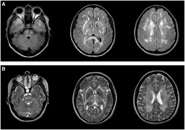
Figure 2.
CADASIL and CARASAL imaging appearance. (A) Typical imaging appearance of CADASIL in axial FLAIR MRI images. There is symmetric subcortical high signal in the anterior temporal lobes, internal and external capsules and scattered asymmetric involvement of the periventricular cerebral white matter and pons. (B) CARASAL in T2 axial images. There was no involvement of the anterior temporal lobes (left) but there was extensive involvement of the internal and external capsules, the basal ganglia and thalami (middle) and the periventricular and deep white matter (right).

